Our laboratory is broadly focused on developing new molecules and materials for molecular imaging and drug delivery. Recent projects in the lab include the development of new probes for in vivo imaging of bacterial infections and reactive oxygen species. In addition, we also have a long standing interest in developing new materials for intracellular protein delivery.
Molecular Imaging
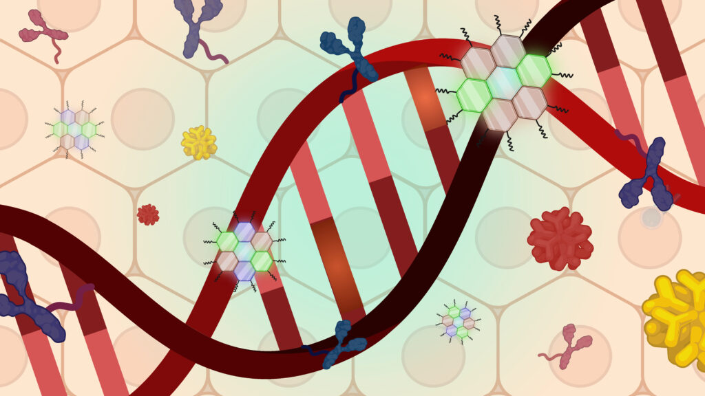
The Murthy laboratory has several projects in the area of molecular imaging. For example, we are developing probes that can image reactive oxygen species and bacterial infections. We are interested in using these new probes for in vitro diagnostics and in vivo imaging applications.
Hydrocyanines
In 2009 the Murthy laboratory developed a new family of fluorescent probes that can image superoxide and the hydroxyl radical in cell culture, in tissue and for the first time in vivo. The development of fluorescent probes for superoxide and the hydroxyl radical is a central problem in the field of chemical biology. Superoxide and the hydroxyl radical play a significant role in a variety of inflammatory diseases and probes that can detect these reactive oxygen species (ROS) have tremendous potential as medical diagnostics and research tools. Fluorescent sensors for superoxide and the hydroxyl radical, such as dihydroethidium (DHE), have been developed, however, they have had limited applicability due to their spontaneous auto-oxidation, rapid photobleaching, low emission wavelengths and multiple reaction products with ROS. New chemical probes for superoxide and the hydroxyl radical are therefore greatly needed. To address this need, the Murthy laboratory developed a new family of fluorescent ROS sensors, termed the hydrocyanines, which can be synthesized in one step from commercially available cyanine dyes, and can detect superoxide and the hydroxyl radical in living cells, serum, tissue samples and in vivo. We are currently collaborating with approximately 30 laboratories on using hydrocyanines for various applications and anticipate widespread interest in the hydrocyanines given their unique physical/chemical characteristics. The first paper on the hydrocyanines was published in Angewandte Chemie International, several others are currently pending. The hydrocyanines are now commercially available from Licor Biosciences, under the name ROSstar.
Kundu K, Knight S, Willett N, Lee S, Taylor W, Murthy N. “Hydrocyanines: A new class of fluorescent sensors that can image reactive oxygen species in cell culture, tissue and in vivo”. Angewandte Chemie International (2009), 48(2), 299-303.
Lin P, Myers L, Ray L, Song S, Nasr T, Berardinelli A, Kundu K, Murthy N, Hansen J, Neish A. “Lactobacillus rhamnosus blocks inflammatory signaling in vivo via reactive oxygen species generation”. Free Radical Biology & Medicine (2009), 47(8), 1205-1211.
Maltodextrin-based Bacterial Imaging Agents
Bacterial infections causes millions of deaths each year and new strategies for improving their treatment are greatly needed. One of the major challenges in treating bacterial infections is diagnosing them at an early stage. At present bacterial infections can only be diagnosed after they have caused significant tissue damage, or via a blood culture, however both of these methods can only diagnose late stage infections, which are challenging to treat because of the high bacterial burden. There is therefore a great need for developing new strategies to diagnose bacterial infections
We have developed a strategy to image bacterial infections based on targeting the maltodextrin transporter. We identified the maltodextrin transporter as a receptor for targeting bacteria because it is unique to bacteria and not expressed on mammalian cells, internalizes maltodextrins at a very fast rate and also tolerates large structural modifications of its substrates, allowing us to deliver a wide variety of molecules to bacteria. Using this strategy, we have developed a method to deliver imaging and therapeutic agents to bacteria.
Ning X, Lee S, Wang Z, Kim D, Subbtlefield B, Gilbert E, Murthy N. “Maltodextrin based imaging probes detect bacteria in vivo with high sensitivity and specificity”. Nature Materials. (2011), 10(8), 602-7
Drug Delivery
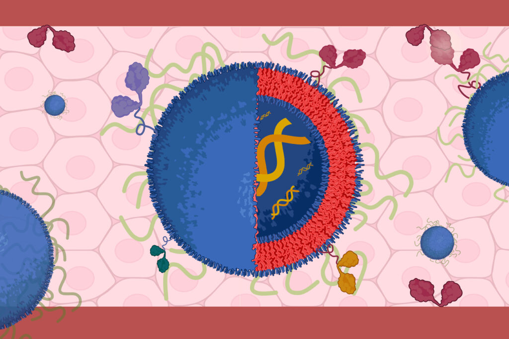
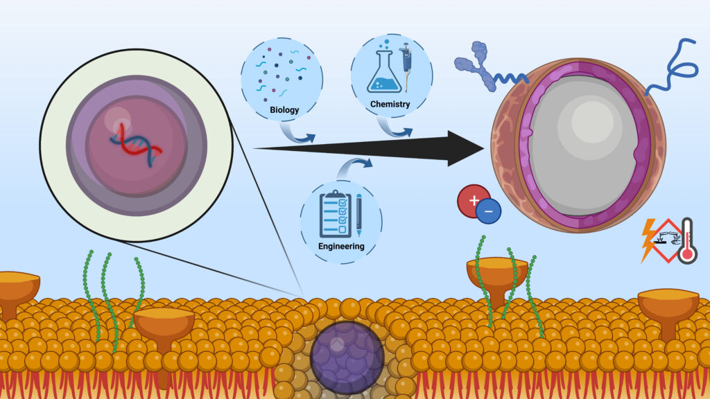
Drug delivery has the potential to transform medicine, due to the great power of biotherapeutics and the delivery challenges preventing them from being effective. Advances in molecular biology during the past twenty years have generated thousands of potential new therapeutics, composed of proteins, RNA and DNA, which have the potential to be significantly more effective than conventional small molecule drugs (biotherapeutics). However, delivery challenges have limited the efficacy of these new therapeutics, and our lab is therefore focused on developing new delivery vehicles that can enhance the efficacy of RNA, DNA and protein based drugs.
Polyketals
Our laboratory has developed a new family of polymers for drug delivery, termed the polyketals. The polyketals are synthetic polymers with ketal linkages in their backbone, and have enormous potential for drug delivery because of their acid sensitivity and biocompatible degradation products. Polyketals had never been synthesized, prior to 2005, because of a lack of synthetic methodologies. In 2005, the Murthy laboratory demonstrated that it was possible to synthesize polymers with ketal linkages in their backbone, using a synthetic procedure based on the acetal exchange reaction. This synthetic procedure generates multigram quantities of polyketals in one step, can easily be scaled up for industrial applications, and has now been used to generate numerous polyketals.
Polyketal microparticles have shown great promise for the treatment of a variety of inflammatory diseases, in preclinical, animal models of disease. Polyketal microparticles hydrolyze rapidly at the acidic pH of the macrophage phagosome, and this property has allowed polyketals to be effective in delivering therapeutics to macrophages. For example, polyketal microparticles, 1-3 microns in size, have been used to deliver siRNA, proteins and small molecule therapeutics to macrophages in mice, and enhance the treatment of acute liver failure and acute lung injury. Large drug loaded polyketal microparticles, 10-20 microns in size, are also being investigated as injectable controlled release reservoirs, and have been able to enhance the treatment of myocardial infarcts and arthritis in mice and rats. Several papers have been published on the polyketals, a few of these are listed below. In addition, three patent applications (one international) have been filed on the polyketals, and a company, Ketal Biomedical Inc., has been formed to translate the polyketals into clinical trials.
Heffernan M, Murthy N. “Polyketal nanoparticles: A new pH sensitive, biodegradable drug delivery vehicle”. Bioconj. Chem. (2005), 16(6), 1340-1342.
Khaja S, Lee S, Murthy N. “Acid degradable protein delivery vehicles based on metathesis chemistry”.Biomacromolecules (2007), 8(5), 1391-1395.
Lee S, Yang SC, Heffernan MJ, Taylor WR, Murthy N. “Polyketal microparticles: a new delivery vehicle for superoxide dismutase”. Bioconj. Chem. (2007), 18(1), 4-7.
Yang SC, Bhide M, Crispe IN, Pierce RH, Murthy N. “Polyketal Copolymers: A new acid-sensitive delivery vehicle for treating acute inflammatory diseases”. Bioconj. Chem. (2008), 19(6), 1164-1169.Sy J, Iyer G, Yang S, Brown M, Oh T, Dikalov S, Murthy N, Davis ME. “Sustained release of a p38-inhibitor from non-inflammatory microspheres inhibits cardiac dysfunction”. Nature Materials (2008), 7(11), 863-868.
Polythioketals
Inflammatory bowel disease (IBD) affects millions of people each year and effective treatments are greatly needed. The overproduction of TNF-α is a central cause of IBD, and there is therefore great interest in developing IBD therapeutics that can inhibit TNF-α. However, TNF-α is also needed for a functioning immune system and systemically inhibiting TNF-α causes toxic side effects. Thus, therapeutic strategies are required to selectively inhibit TNF-α in inflamed mucosal tissues.
We developed a family of particles, termed the polythioketal particles, that are designed to deliver siRNA orally and target inflamed intestinal tissues. Polythioketals are designed to be stable in the acidic environment of the stomach and to enzymatic degradation in the intestines, but are degradable in the presence of reactive oxygen species, which are generated during inflammation. Using this strategy, we delivered siRNA to inhibit TNF-α in inflamed intestinal issues.
Wilson DS, Dalmasso G, Wang L, Sitaraman SV, Merlin D & Murthy N. ”Orally delivered thioketal nanoparticles loaded with TNF-α–siRNA target inflammation and inhibit gene expression in the intestines” Nature Materials, (2010), 9, 923-928.
Direct Reprogramming of Cardiac Fibroblasts into Cardiomyocytes
Functional recovery of damaged cardiac muscle after MI remains challenging due to the limited regeneration capacity of cardiomyocytes. One powerful approach to cardiac regeneration is direct reprogramming of cardiac fibroblasts into cardiomyocyte-like cells with transcription factors (TFs) as fibroblasts comprise more than 60% of all the cardiac cells, which provide a potential source for cardiomyocytes. However, current methods of direct reprogramming via viral transfection face significant hurdles in a clinical context due to the potential risks of immune response and viral gene integration, which may lead to cancer or diseases. Therefore, there is a great need for the development of new technologies that can deliver TFs, and we are currently developing transcription factor delivery vehicles for direct reprogramming.